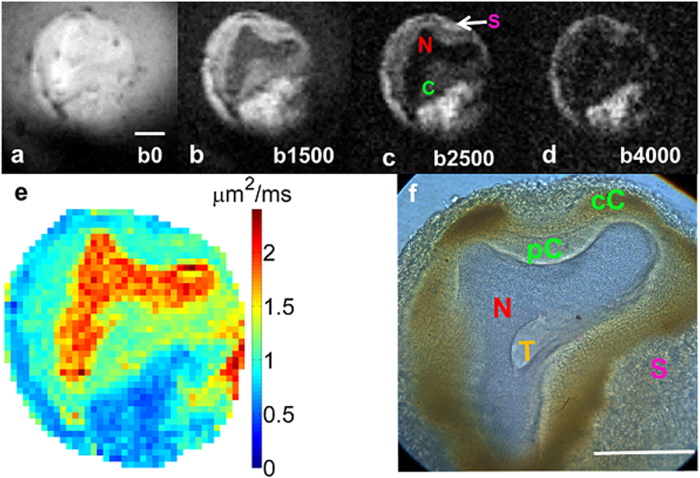
Figure 5. Diffusion weighted images of a slice of the single neuron embedded in agarose.
MRM scan parameters: TE/TR = 20/2000 ms, resolution = 11.7 × 11.7 × 100 μm3, NEX = 28, acquisition Time = 2 hours, bandwidth = 50 kHz, read gradient amplitude = 784 mT/m, and phase gradient amplitude = 743 mT/m, b-values = (a) 0, (b) 1500, (c) 2500, (d) 4000 s/mm2 (e) ADC map in mm2/s, diffusion gradient separation (Δ)/ diffusion gradient duration (δ) = 11 ms/1 ms. (f) Light microscopy image of the same neuron showing five distinct subcellular tissue types at 40x magnification. Of particular note is the morphological delineation of more than two intracellular compartments consisting of nucleus (N), perinuclear cytoplasm (pC), cortical cytoplasm (cC) enriched with mitochondria and lipochondria functioning as photoreceptors, and possibly trophospongium (T), which is glial contact to the large neuron via membrane invagination. Scale bar is 100 μm.

