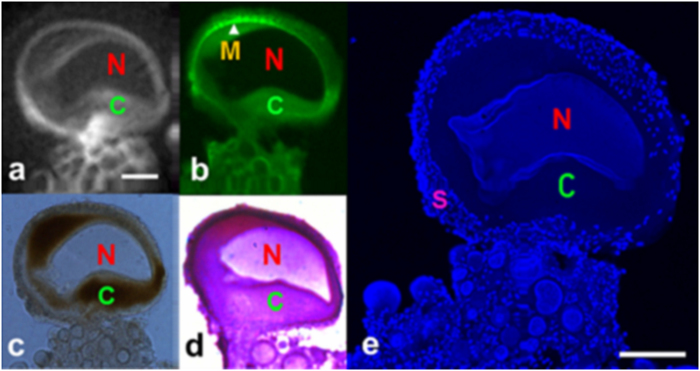
Figure 6. Direct correlation of cellular architecture of single neurons of Aplysia californica in MRM and histological images.
(a) 2D diffusion-weighted images at the in-plane resolution of 7.8 μm2 with 50 μm thick slice at b = 1500 s/mm2. (b) Green fluorescent colored PKH 67-stained plasma membrane. (c) Bright-field microscopy. (d) Nissl-stained cytoplasmic region of the adjacent slide. (e) DAPI-stained nucleic acid in the nucleus in a single neuron; smaller cells and numerous tiny satellite cells around the periphery of the neuron. MR scan parameters: TE/TR = 20/2000 ms, resolution = 11.7 × 11.7 × 150 μm3, NEX = 100, FOV = 1.5 × 1.5 × 0.15 mm, acquisition time = 2 hours, b-values = 1500 s/mm2, diffusion gradient separation (Δ)/diffusion gradient duration (δ) = 5 ms/1 ms. Nucleus (N), cytoplasm (C), plasma membrane(M), and satellite cells(S). Scale bar is 100 μm.

