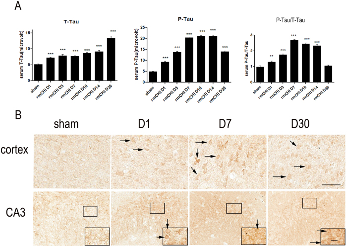
Figure 5. Tau and phosho-Tau changes after repetitive closed head injury.
(A) Detection of Tau by a-EIMAF in mouse serum following rCHI. Mice were subjected to repetitive mTBI and blood was collected at various time points as indicated. The levels of T-Tau (left) and P-Tau (middle) as well as the ratio of P-Tau to T-Tau (right) in serum were determined by a-EIMAF. Statistical analysis (t-test) was based on comparison to naïve: ** p < 0.01; *** p < 0.0001. (B) Immunostaining of mice brain sections using P-Ser202 (CP13) antibody to assess changes in tau. Representative images obtained from mouse brain sections showing P-Tau immunoreactive cells in the cortex and hippocampal CA3 pyramidal neurons at different time points post-rCHI. The insert show magnification of the dense P-Tau stained cells in each section. Scale bar = 50 μm. Arrows indicated representative P-Tau positive cells in each section.

