
Figure 3.
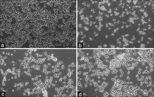
293T cells were seeded in 12-well plates (50,000 cells/well) and the number of live and dead cells was determined by the tryphan blue exclusion test and the cells were examined at the end of 72 h. Appearances of the cells untreated (A: Control) and treated (B: Doxorubicin-HCl, C: Aerial extract, D: Root extract) were photographed under a contrast phase microscope (Olympus-IX71)