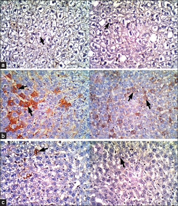
Figure 1.

Immunohistochemical staining of the liver sections (×400) stained with caspase 3 antibodies for apoptotic cells and hematoxylin counterstaining of the nuclei and non-apoptotic cells. (a) control groups, (b) CCl4 groups and (c) rats received ethanolic extract of Schinus terebinthifolius either before [left side pictures] or after [right side pictures] CCl4-intoxications. Arrowheads in figures (b) point out caspase-3 positive apoptotic cells
