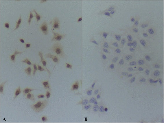
Fig. 2.

HPV L1 immunocytochemistry assay. a HeLa cells. There were positive reactions which were yellow in cytoplasm and nucleus in HeLa cells detected using anti-HPV L1 monoclonal antibody and the stain in cytoplasm is darker. The reaction shows the existence of HPV L1 in HeLa cells. b HaCat cells. No positive reaction was detected in HaCat cells using anti-HPV L1 monoclonal antibody
