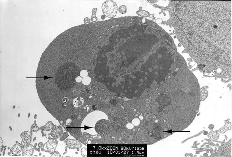
Fig. 3.

HeLa cell electron micrograph. In the graph, there is a HeLa cell in apoptosis. Many clumped inclusion bodies can be seen in the HeLa cell cytoplasm (indicated by the arrow). These clumped bodies appear in different sizes and shapes. No inclusion bodies are seen in the cell nucleus
