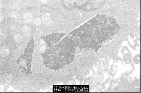
Fig. 6.

The graph of partial enlargement of the HeLa cell electron micrograph in Fig. 5. Two of the inclusion body structures are highly magnified. At higher magnification, this inclusion body can be seen as a dense aggregation of plenty of tiny granules with uniform diameter
