Figure 1.
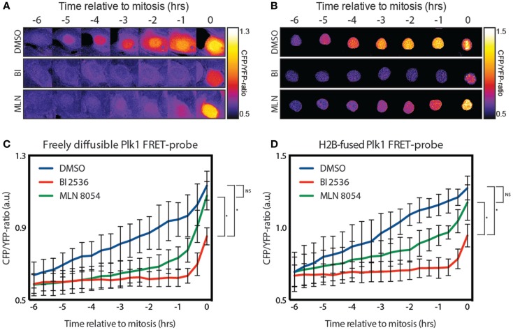
Plk1 activity is first seen in the nucleus. (A) Stills from a movie showing false color-coded CFP/YFP emission ratios. The stills show control-, BI 2536-, and MLN 8054-treated U2OS cells expressing the diffusible FRET-based biosensor for Plk1 activity while entering mitosis. BI 2536 was used at a concentration of 100 nM; MLN 8054 at a concentration of 1 μM. (B) Stills from a movie showing false color-coded CFP/YFP emission ratios of U2OS cells expressing the H2B-tagged FRET based biosensor were treated as in (A). (C) Quantification of CFP/YFP-ratio of cells shown in (A). Error bars represent the SD of 10 individual cells. (D) Quantification of CFP/YFP-ratio of cells shown in (B). Error bars represent the SD of 10 individual cells; *p < 0.0001.

