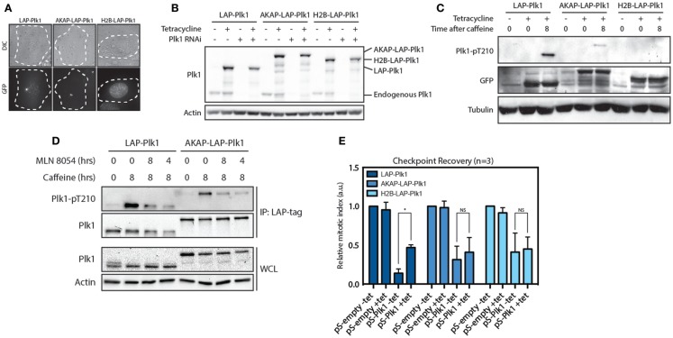
Figure 2.
Plk1 is activated at the centrosomes but needs to be dynamically localized. (A) Expression of RNAi resistant LAP-Plk1, AKAP-LAP-Plk1 or H2B-LAP-Plk1 was induced in U2TR stably expressing these constructs by addition of tetracycline. DIC- and GFP-images were taken of representative cells. (B) Tetracycline inducible U2TR cells stably expressing RNAi resistant LAP-Plk1, AKAP-LAP-Plk1 or H2B-LAP-Plk1 were transfected with an empty pSuper, or a pSuper targeting endogenous Plk1. Cells were synchronized in G2 and damaged with 0.5 μM adriamycin for 1 h and expression was induced where indicated using tetracycline. 16 h after induction of DNA damage cell were harvested and analyzed by western blotting. (C) Tetracycline inducible U2TR cells stably expressing RNAi resistant LAP-Plk1, AKAP-LAP-Plk1 or H2B-LAP-Plk1 were synchronized in G2 and damaged with 0.5 μM adriamycin for 1 h and expression was induced where indicated using tetracycline for 16 h. Cells were harvested at the indicated time points after addition of caffeine and analyzed by western blotting. (D) Cells were treated as in C. MLN 8054 was added at a concentration of 1 μM either for 8 h together with caffeine or during the last 4 h of caffeine. LAP-tagged proteins were immunoprecipitated with S-protein agarose beads and analyzed by western blot. (E) Tetracycline inducible U2TR cells stably expressing RNAi resistant LAP-Plk1, AKAP-LAP-Plk1 or H2B-LAP-Plk1 were transfected with an empty pSuper, or a pSuper targeting endogenous Plk1. Cells were synchronized in G2 and damaged with 0.5 μM adriamycin for 1 h and expression was induced where indicated using tetracycline. Cells were arrested for 16 h, recovery was induced by caffeine addition for 8 h and the mitotic index was determined, based on the percentage of Histone H3-pS10 positive cells, using FACS. Error bars represent the SD of three independent experiments; *P < 0.001.

