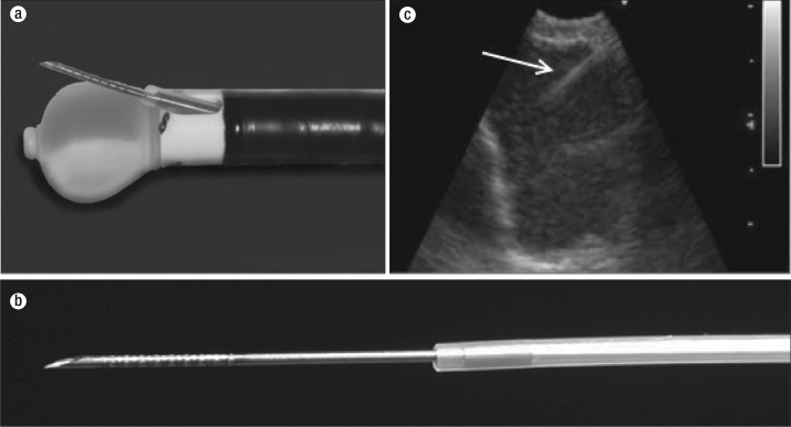
Figure 3.
Endobronchial ultrasound (EBUS) bronchoscope. (a) The inflated balloon over the ultrasound transducer and a protracted biopsy needle through the EBUS working channel. (b) An EBUS biopsy needle with dimples for better sonographic visualization (arrow). (c) Real-time visualization of EBUS transbronchial aspiration needle advancement into the lymph node.

