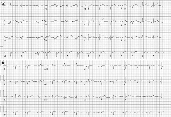
Figure 1.
Electrocardiograms in case 1. (a) Initial electrocardiogram demonstrates an ectopic atrial rhythm with an isolated sinus beat (third beat), inferior Q waves, and ST elevation in the inferior leads. (b) Repeat electrocardiogram shows Q waves in the inferior leads and resolution of inferior ST elevations. There is limb lead reversal.

