Figure 3.
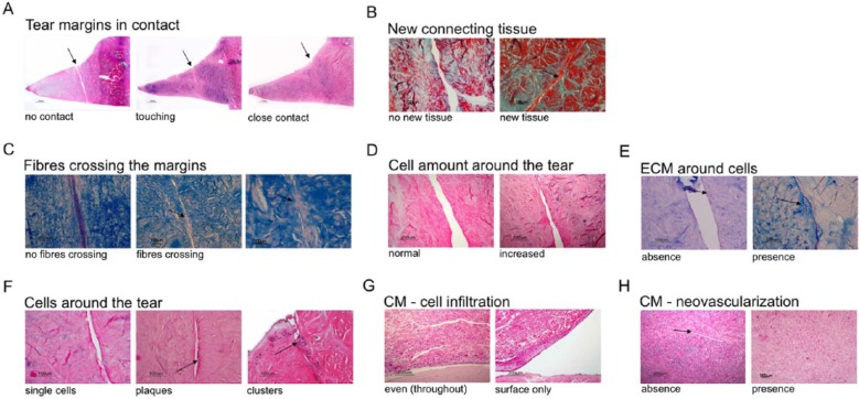
Histology scoring approach. Parameter “tear margins in contact” (A) distinguished between “no contact,” “touching,” and “close contact,” where “touching” and “close contact” were considered as indicative of healing (H&E); parameter “new connecting tissue” (B) evaluated the absence or presence of new tissue (Masson Trichrome); parameter “fibers crossing the tear” assessed absence or presence of fibers bridging the two tear margins (Alcian blue); parameter “cell amount around the tear” (D) evaluated no increase (normal) or increase in cell numbers around the tear compared to the surrounding meniscus tissue (H&E); parameter “ECM around cells” (E) evaluated the absence or presence of extracellular matrix proteoglycans produced by cells (Alcian blue); parameter “cell organizations around the tear” (F) distinguished between single cells and groups of cells consisting of clusters and/or plaques (H&E); parameter “CM cell infiltration” (G) assessed cellular infiltration of either only the surface or throughout (even) the entire collagen membrane (H&E); parameter “CM neovascularization” (H) evaluated the absence or presence of blood vessels (indicated with an arrow) within collagen membrane (H&E stain). H&E = hemotoxylin–eosin.

