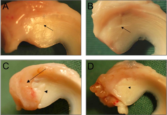
Figure 5.
Macroscopic evaluation of tear appearance and tear coverage with collagen membrane. Tears in contact were considered as either healed, with visible connecting tissue between the margins (A), or not healed, with a visible gap between the margins (B), indicated with arrowheads. The partial or complete degradation of collagen membrane allowed for partial tear coverage (C), indicated with an arrow, or complete tear exposure (D).

