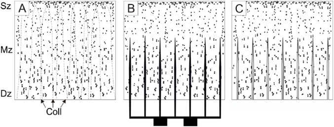
Figure 2.

A schematic drawing to illustrate the production of multiply incised pure chondral graft. Dots represent lacunae, the 3 zones of hyaline cartilage are labeled Sz (superficial zone), Mz (middle zone), and Dz (deep zone). (A) Cross-section of the full-thickness articular cartilage specimen harvested from a donor surface. Collagen fiber pattern is shown with broken lines (Coll). (B) Incision of the graft using multiple parallel blades. The cuts go through the deep and middle zone of cartilage but leave the superficial zone uncompromised. Please note, the incisions run parallel with the collagen fibers (Coll), thus, collagen network is left considerably intact. (C) Processed chondrograft ready to be implanted.
