Figure 3.
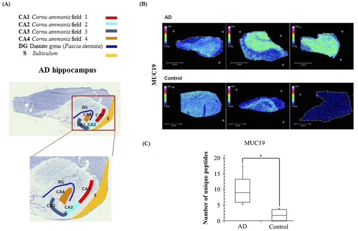
(A) Annotated optical image of a hippocampal section obtained from an Alzheimer’s disease patient stained with toluidine blue; Horn of Ammon (CA), dentate gryus (DG), and subiculum (S). (B) MUC19 isoform 5 is highly expressed in AD hippocampal tissues. MALDI imaging mass spectrometry (MSI) data (MUC19; m/z 11,135 ± 0.05%). (C) Spectral counting data for MUC19.

