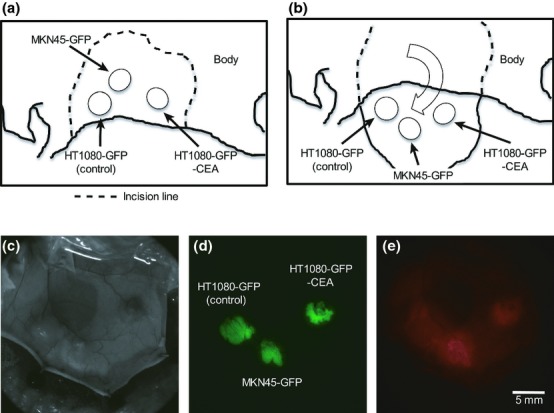
Figure 2.

Inoculation of human cancer cells into immunodeficient mice and in vivo macroscopic imaging using a fluorescence zoom microscope. (a) Schema of the sites of inoculation of human cancer cells. The cells were inoculated s.c. into the back skin of nude mice at the rostral–ventral site (HT1080-GFP cells), the caudal–ventral site (HT1080-GFP-CEA cells) or the dorsal site (MKN45-GFP cells). (b) Schema of preparation of skin flaps. Seven or eight days after the inoculation, the inoculation sites were exposed by the skin-flap method. (c–e) In vivo macro imaging of tumors. In vivo macro imaging of the tumor masses was performed using a fluorescence zoom microscope 24 h after injection of Alexa Fluor 594-conjugated anti-CEA antibody (50 μg/mouse). Exposure times for the GFP and Alexa Fluor 594 fluorescence images were 30 and 100 ms, respectively. These experiments were repeated three times and similar results were obtained.
