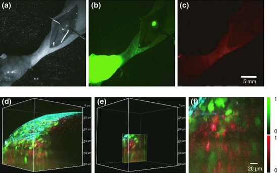
Figure 5.

In vivo fluorescence macroscopic and microscopic imaging of lymph-node metastases by a fluorescence zoom microscope and a two-photon excitation microscope. (a–c) A footpad spontaneous metastasis model using HT1080-GFP-CEA cells observed by a fluorescence zoom microscope. The popliteal lymph node was exposed, and multiple images were collected: bright field image (a), GFP (b) and Alexa Fluor 594 (c). Exposure times for the GFP and Alexa Fluor 594 images were 1000 and 3000 ms, respectively. (d–f) Two-photon excitation microscopy of the popliteal lymph node. After in vivo macroscopic imaging, the same lymph node was observed using a two-photon excitation microscope. Acquired images are shown as 3-D construction (d), cropped 3-D image of Figure 5d (e) and magnified image of Figure 5e (f), respectively. Red, green and blue indicate Alexa Fluor 594 fluorescence, GFP fluorescence and second harmonic generation (SHG), respectively.
