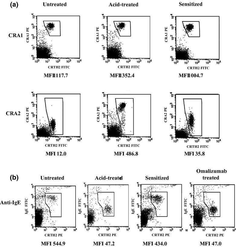
Fig 2.

Binding levels of CRA1, CRA2, and anti-IgE antibody measured by basophil staining to confirm the passive sensitization by flow cytometric analysis. (a) Immunostained healthy subject's basophils with anti-FcεRI antibodies; PBMCs with or without acid treatment were stained for CD3, prostaglandin D2 receptor (CRTH2), and FcεRI. (b) Immunostained healthy subject's basophils with anti-IgE antibody; PBMCs with or without acid treatment were stained for CD3, CRTH2, and IgE. Flow cytometer charts for CD3− PBMCs are shown. Basophil fraction (CD3− and CRTH2+ PBMC) was gated. MFIs indicated for binding levels of anti-CRA1, anti-CRA2, and anti-IgE antibodies on basophils. MFI, mean fluorescence intensity.
