
Fig 1.
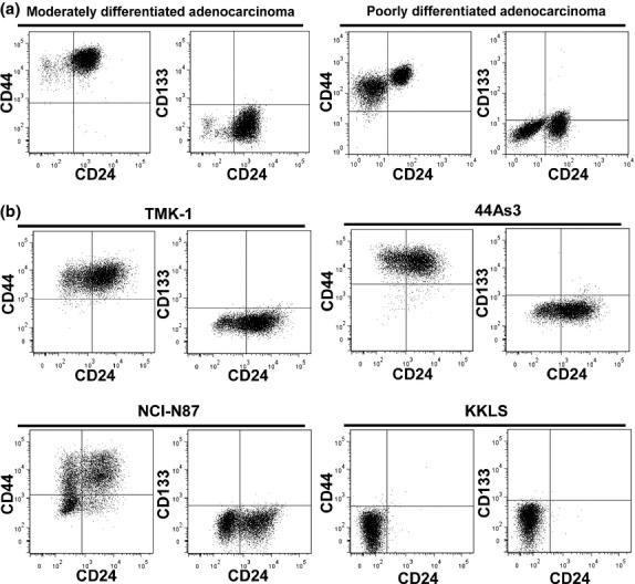
Fluorescence-activated cell sorting (FACS) analyses of representative cell surface markers in GCa cells. (a) Human primary tumor cells collected from ascites of patients. Left, moderately differentiated adenocarcinoma; right, poorly differentiated adenocarcinoma with signet ring cell. (b) GCa cell lines. Top left, TMK-1; top right, 44As3; bottom left, NCI-N87; bottom right, KKLS.