
Fig 1.
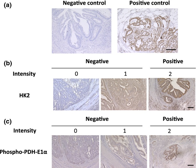
Immunohistochemical analysis of HK2 and p-PDH in clinical colorectal cancer samples. (a) Phosphate buffered saline was used as a negative control and a case of pancreatic cancer tissue was used as a positive control for HK2. (b) Staining of HK2 and (c) p-PDH at the invasive front are shown; the intensity was rated in three stages. Scale bar, 200 μm.