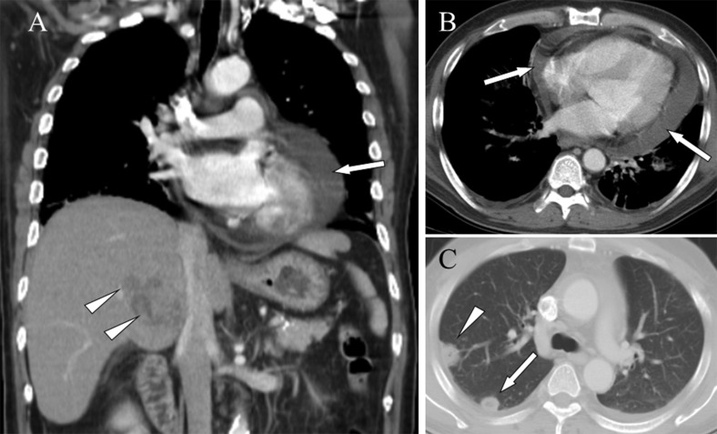
Figure 5.
Pericardial effusion and septic pulmonary embolism caused by a Klebsiella pneumoniae liver abscess in a 73-year-old woman. A) A mediastinum window of a coronal computed tomography scan reveals a hypodense, hypovascular mass of approximately 5 cm in diameter in the S7 area of the right hepatic lobe (arrowheads) and fluid in the pericardial space (arrow). B) A mediastinum window of a cross-sectional computed tomography scan shows fluid in the pericardial space (arrows). C) A lung window of a cross-sectional computed tomography scan shows a peripheral wedge-shaped (arrowhead) and a peripheral nodular (arrow) opacity.

