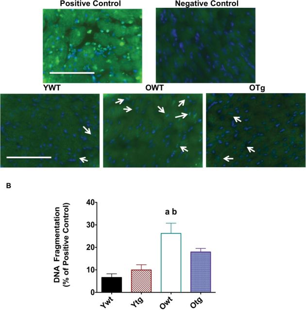
Figure 7.
Terminal deoxynucleotidyl transferase-mediated dUTP nick-end labeling-positive staining (A) is visualized by immunofluorescence staining (×40) in left ventricle (LV) samples of young adult wild-type (YWT), old WT (OWT), and old Sod2 Tg (OTg), and DNA fragmentation (B) was used as a marker of apoptosis through quantification of mononucleosomes and oligonucleosomes via ELISA in LV samples of YWT, young adult Sod2 Tg (YTg), OWT, and OTg. Data are expressed as means ± standard error of the mean. “a” indicates a significant difference vs YWT (p < .05). “b” indicates a significant difference vs YTg (p < .05).

