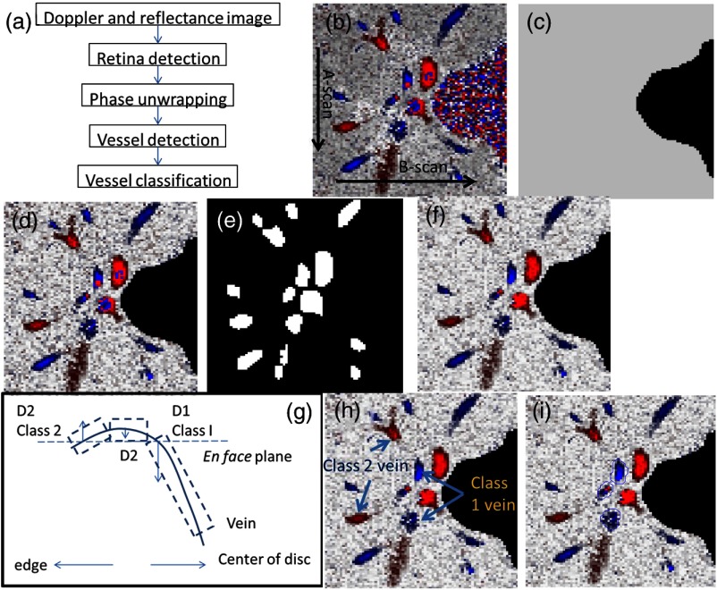
Fig. 5.
Detection of veins in Doppler OCT image. (a) Flow chart for vessel detection; (b) Doppler phase shift and reflectance Image on one en face plane. The arrow is the direction of B-scans and A-scans. (c) Binary mask include retina; (d) masked Doppler OCT image using retinal mask; (e) vessel mask in one en face plane after vessel detection; (f) Doppler OCT image in Fig. 5(e) after phase unwrapping; (g) illustration of two types of vein classification rules: vein (class 1) is the vein inside the disc, vein (class 2) is the vein outside the disc. At depth showed in this figure, the en face plane cross with the vein twice; (h) two type of veins in same en face plane; and (i) veins (class 1) detected in one en face plane (blue circle).

