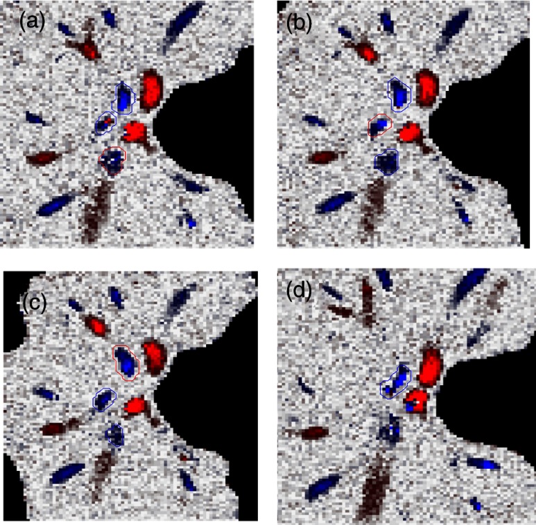
Fig. 6.
Illustration of multiple plane en face Doppler method. (a)—(c) Three en face planes were selected for comparison. Each vein had an optimized en face plane in which the Doppler phase shift was strong enough but without any artifact. Blue circle is the valid vein mask. Red circle means the particular vessel obtained higher flow measurement than other planes. (d) When two branches merged to one vessel in deeper en face plane, the summation of branches [indicated by the upper two circles in Fig. 6(a)] will be compared to the flow in the merged vessel [indicated by the blue circle in Fig. 6(d)].

