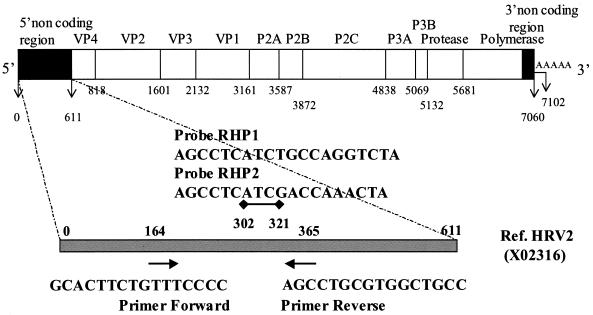
FIG. 1.
Schematic representation of the rhinovirus genome. The locations and orientations (arrows) of the primers and the two probes used in the real-time RT-PCR are indicated. Initiation and cleavage sites are defined by the vertical bars, their positions, and the proteins coded for by the reference HRV-2 isolate. The gray bar represents the 5′ NCR from which the consensus sequence, the primers, and the probes were defined.

