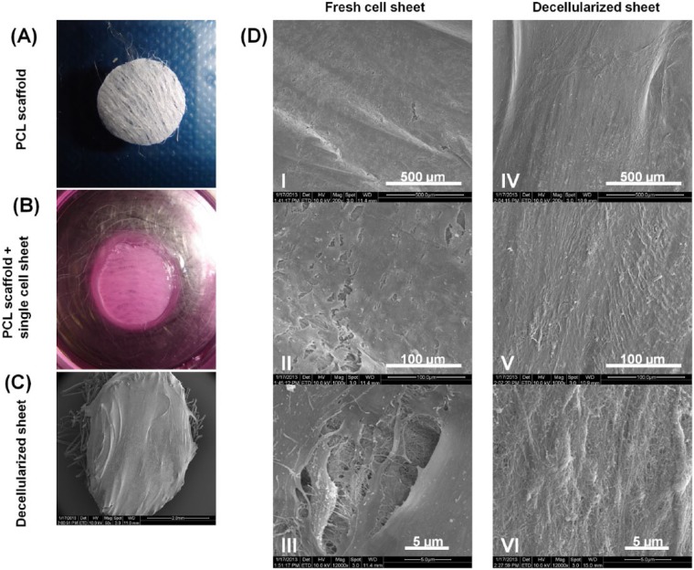
Figure 1.
Harvesting of fresh human periodontal ligament (HPDL) cell sheet and scanning electron microscopy (SEM) showing fresh and decellularized PDL sheet. (A) PCL scaffold 5 mm in diameter, after NH4OH treatment. (B) Fresh HPDL cell sheet attached to sterile PCL scaffold. (C) SEM image of decellularized PDL sheet on top of a PCL scaffold. (D) Different SEM magnification of fresh (I-III) and decellularized (IV-VI) sheets. This figure is available in color online at http://jdr.sagepub.com.

