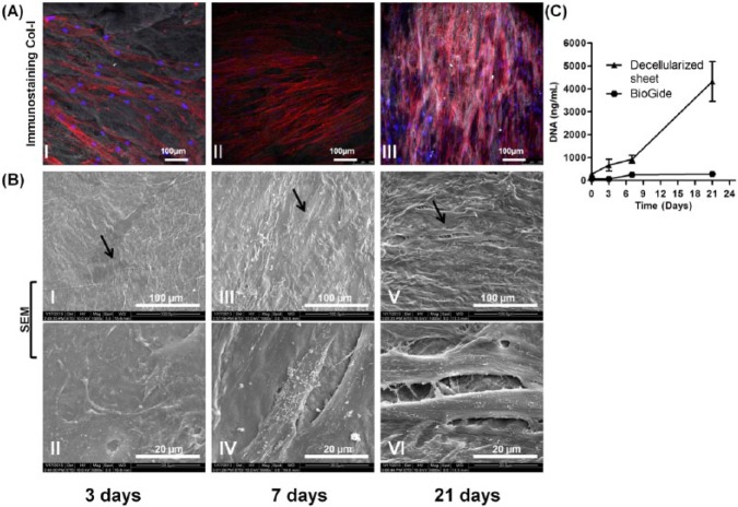
Figure 4.
Recellularization potential of the decellularized sheet after being seeded with allogenic hPDL cells after 3, 7, and 21 days. (A) Confocal imaging and immunostaining of human collagen type I (gray), nuclei (blue), and actin filaments (red). (B) SEM showing hPDL cells at different time points. (C) DNA quantification showing cell proliferation on decellularized constructs and Bio-Gide® membrane over 21 days. This figure is available in color online at http://jdr.sagepub.com.

