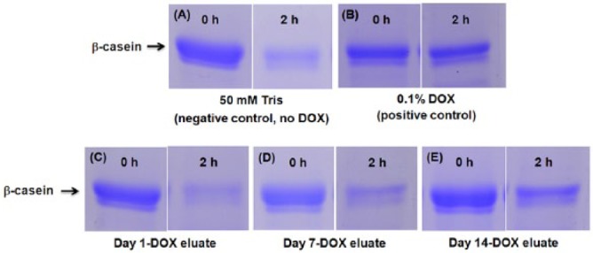
Figure 4.

Representative images of Coomassie blue–stained sodium dodecyl sulfate–polyacrylamide gel electrophoresis gels of β-casein cleavage assays. (A) Negative control: 50 mM Tris with 0.2 M NaCl, 10 mM CaCl2, and 1 µM ZnCl2 (pH 7.4). (B) Positive control: 0.1% doxycycline (DOX) solution (Sigma). DOX eluates: (C) day 1, (D) day 7, and (E) day 14. As depicted, more β-casein is present in panel E compared to the negative control (50 mM Tris) at 2 hr, which is not seen in day 1 and 7 aliquots.
