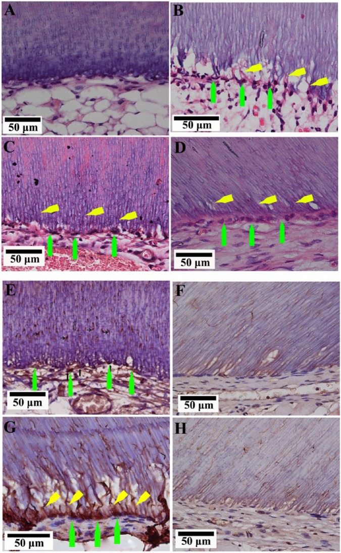
Figure 3.
Regenerated pulp-like tissue with odontoblast-like cells. (A) Empty root slice filled with host adipose tissue did not show an odontoblast-like cell lining. (B) DPSC-alone. (C) DPSC:HUVEC 3:1. (D) DPSC:HUVEC 1:1 microtissue transplanted groups showed odontoblast-like cells (green arrows) along the dentin, projecting their processes (yellow arrows) into the dentinal tubules. Immunohistochemistry for nestin (E, F - negative control) and DSP (G, H - negative control). Green arrows indicate the positively immunostained odontoblast-like cells, and yellow arrows indicate their processes.

