
Figure 1.
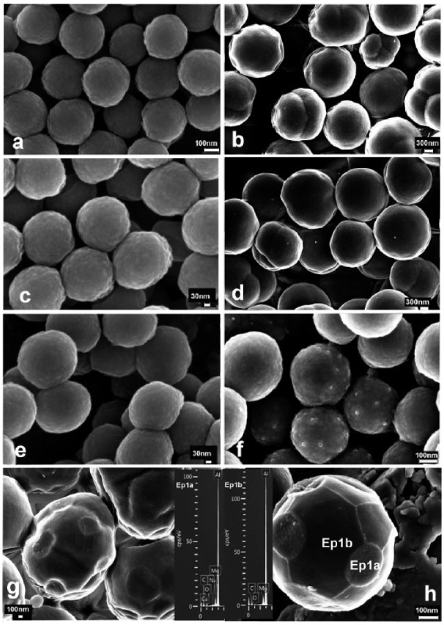
Scanning electron microscopy (SEM) micrographs of nanoparticles (NPs) and microparticles. (a) NPs before immersion in simulated body fluid solution (SBFS) were spherical, around 350 nm in diameter. NPs did not agglomerate. (b) Microparticles before immersion in SBFS were 2.5 µm in diameter. Some concentric, more dense subsurface structures were found. (c) NPs after immersion in 0.1 w/w% ZnCl2 for 3 hr. (d) Microparticles after immersion in 0.1 w/w% ZnCl2 for 3 hr. (e) NPs after immersion in SBFS for 7 days. (f) Zn-NPs after 7 days’ immersion in SBFS. Spotty deposits were distributed throughout nanoparticle surfaces. (g) Microparticles after immersion in SBFS for 7 days. Microparticles showing a heterogeneous composition of central concentric electron reflections and outer increased density are presented. Regularly distributed rounded mineral agglomerates were encountered. (h) Zinc-loaded microparticles after immersion in SBFS for 7 days. Spotty calcium deposits were uniformly distributed throughout particle surfaces, and other formations were grown, forming polygonal lines. Particles lost their regular morphology. Calcium was identified when the spectrum in the energy-dispersive analysis (EDX) was taken from rounded mineral agglomerates (Ep1a), and calcium was not present in these areas (Ep1b). Al and Mg, at the EDX spectra, were contaminant elements from the sample-holder.