Figure 3.

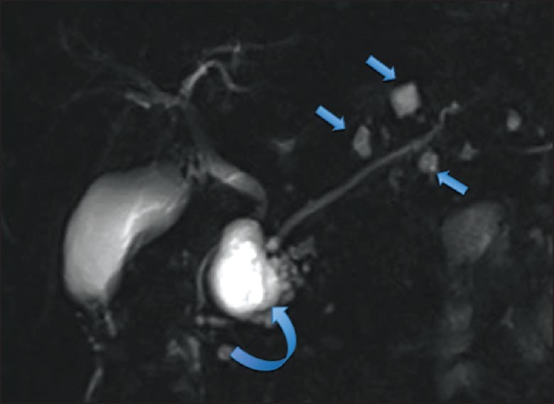
MRI coronal T2 weighted images of BD-IPMN. Multiple cystic dilations of the side branches with the largest lesion in the head (curved arrow) and smaller lesions in the body and the tail (straight arrows)

MRI coronal T2 weighted images of BD-IPMN. Multiple cystic dilations of the side branches with the largest lesion in the head (curved arrow) and smaller lesions in the body and the tail (straight arrows)