
Figure 3.
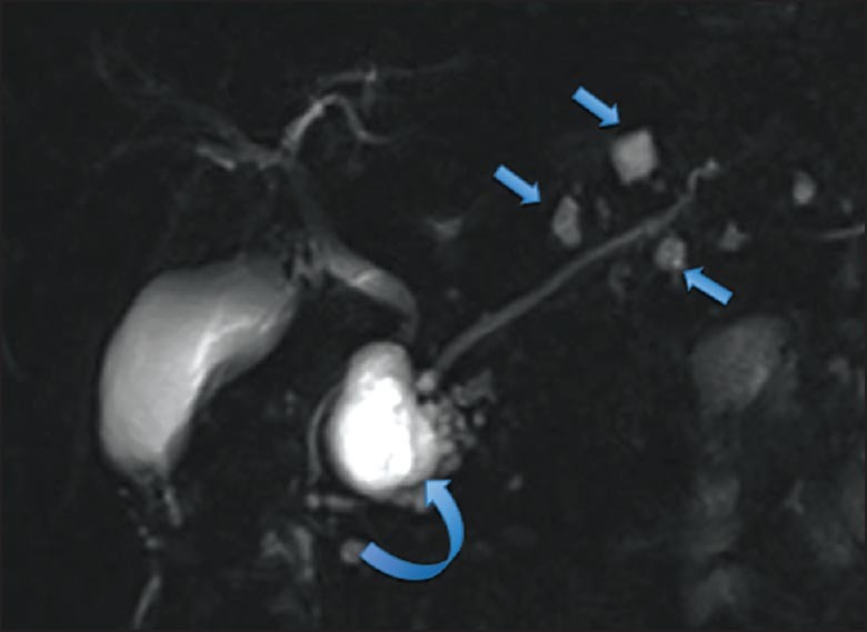
MRI coronal T2 weighted images of BD-IPMN. Multiple cystic dilations of the side branches with the largest lesion in the head (curved arrow) and smaller lesions in the body and the tail (straight arrows)
Official websites use .gov
A
.gov website belongs to an official
government organization in the United States.
Secure .gov websites use HTTPS
A lock (
) or https:// means you've safely
connected to the .gov website. Share sensitive
information only on official, secure websites.
MRI coronal T2 weighted images of BD-IPMN. Multiple cystic dilations of the side branches with the largest lesion in the head (curved arrow) and smaller lesions in the body and the tail (straight arrows)