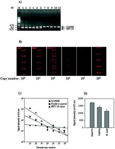
FIG. 4.
Detection limit and reproducibility of the HPV DNA microarray. (A) Agarose gel electrophoresis of the MYH-PCR products with a serial dilution of the plasmid with the HPV-16-specific probe. Ten microliters of each 50-μl PCR product was separated on a 1.0% agarose gel. The input copy numbers of the plasmid with the HPV-16-specific probe are as follows in the indicated lanes: M, 1-kb ladder; 1, 1010 copies; 2, 109 copies; 3, 108 copies; 4, 107 copies; 5, 106 copies; 6, 105 copies; 7, 104 copies; 8, 103 copies; 9, 102 copies; 10, 10 copies; 11, 1 copy. (B) HPV DNA microarray hybridization results with the amplicon obtained by MYH-PCR of the plasmid with the HPV-16-specific probe. Ten microliters of the 50-μl PCR products derived from 105 to 100 copies were hybridized with HPV DNA on the microarray for 2 h at 55°C. The starting plasmid copy number is shown at the bottom. (C) Standard curves of signal intensities for HPV-16 with serial dilutions of 106 to 100 copies. Linear regression was based on the titration series for the plasmid with the HPV-16-specific probe, and each curve is based on the average of three replicates. (D) Reproducibility of the HPV DNA microarray. Three independent experiments were performed with the genomic DNA of Caski cells. The mean ± standard deviation was calculated from the signal intensity of each probe at 635 nm. Each probe is indicated at the bottom: GAPDH, PCR control; Positive, HPV-positive control; HPV-16, HPV-16-specific probe.

