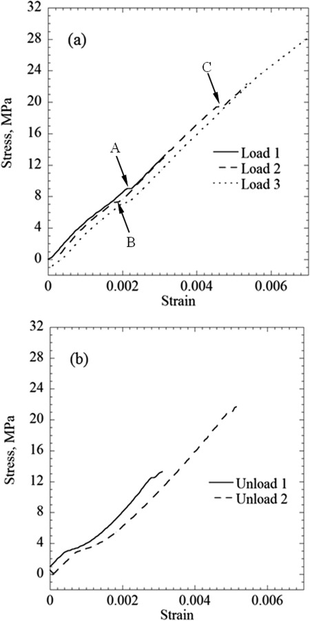
Fig. 7.

Results of repeated loading–unloading cycles of a blood vessel specimen permeated with DP6 + 12% PEG400: (a) the loading portions of the protocol and (b) the unloading portions of the protocol; labels A, B, and C points to steplike changes of strain, possibly associated with the formation of microfractures
