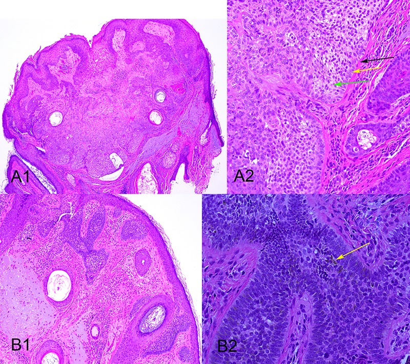
Figure 3A and B.
Dermatopathology images of the lesion shown in figures 1 and 2. A1: Low power view of the trichilemmoma component showing a well-circumscribed, sharply demarcated lesion with surface papillomatosis, three horn cysts and a degree of desmoplasia centrally. A2: Higher power view) shows a thickened basement membrane (black arrow), peripheral palisading (yellow arrow) and clear cells (green arrow). B1: Low power view of the BCC component showing superficial BCC at the dermo-epidermal junction. B2: Higher power view of the BCC with low light, showing melanin pigment (arrow). [Copyright: ©2015 Al Kaptan et al.]

