
FIGURE 4:
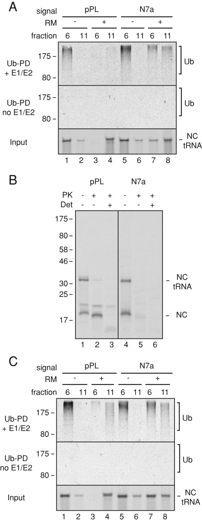
Ubiquitination of membrane-bound RNCs. (A) Short RNCs containing the pPL or N7a signal peptides were produced in reactions containing or lacking RMs. The RNCs were separated by sucrose gradient (as in Figure 3D), and fractions 6 and 11 were subjected to ubiquitination by addition of purified E1, E2, ubiquitin, and ATP. Control reactions lacked E1 and E2. The input RNCs (bottom) and ubiquitin pull downs (Ub-PD) from reactions containing (top) or lacking (middle) E1 and E2 were analyzed by SDS–PAGE and autoradiography. Note that the RNCs in fraction 11 without microsomes appear to be aggregates that are not ubiquitinated (e.g., lanes 2 and 6). (B) Membrane-bound short RNCs prepared as in A were analyzed by a protease-protection assay. Reactions lacked or contained proteinase K (PK) without or with detergent (Det). The tRNA-linked and hydrolyzed nascent chains (NCs) are indicated. The additional higher–molecular weight band in the pPL sample is glycosylated product (Figure 3B). (C) Experiment as in A but using long RNCs of pPL and N7a constructs.