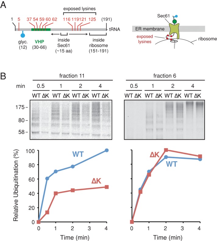
FIGURE 6:
Identification of lysines involved in RNC ubiquitination at the membrane. (A) Schematic of the signal-cleaved pPL long nascent chains labeled as in Figure 3A. The approximate regions inside the ribosome exit tunnel and within the Sec61 channel are indicated by brackets. The arrows depict the potential range of nascent chain sliding within the Sec61 channel. The four lysine residues mutated to arginine to generate the ∆K construct are indicated. Right, hypothesized architecture of the RNC, showing the four lysines (red dots) that may be exposed at the ribosome–translocon junction. (B) Long pPL RNCs or a matched construct lacking lysines 116, 119, 121, and 125 (termed ∆K) were targeted to the membrane, isolated as in Figure 3D, and subjected to a ubiquitination time course. A parallel reaction was performed on the identical cytosolic RNCs (right). The purified ubiquitinated products were analyzed by autoradiography (top), the quantification of which is plotted at the bottom.

