FIGURE 1:
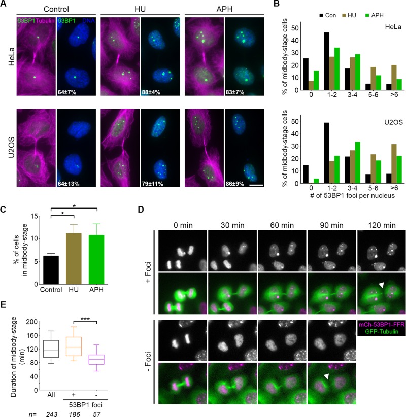
53BP1 foci are present in nuclei before abscission, and their presence corresponds to longer time in midbody stage. (A) HeLa and U2OS cells were cultured for 24 h in the presence of DMSO (control), HU, or APH and analyzed for the presence of nuclear foci containing 53BP1 (green). Midbody-stage cells were identified with an antibody directed against α-tubulin (magenta), and DNA was stained with DAPI (blue). Total percentage of midbody-stage cells with 53BP1 foci after each treatment is indicated (mean and SD from four experiments). Scale bar, 10 μm. (B) Quantification of 53BP1 foci per daughter cell nucleus in midbody-stage cells from each cell line after the indicated treatments. Data are combined results from four experiments. (C) Quantification of the number of midbody-stage cells (HeLa) after the indicated treatments. Error bars are mean and SD from three experiments. *p < 0.05 (Mann–Whitney test). (D) Montages of HeLa cells expressing GFP-tubulin and mCherry-53BP1-FFR subjected to time-lapse imaging. Time from midbody formation (0 min) to midbody disassembly (white arrowheads) was quantified by tracking GFP-tubulin for all cells that passed through this stage, with or without 53BP1 foci. (E) Quantification of abscission timing as defined in D. Boxplot represents the 25th, median, and 75th percentile of values from the indicated treatments (n = number of cells per treatment, combined from four experiments). Whiskers represent the 10th and 90th percentiles. ***p < 0.0001.

