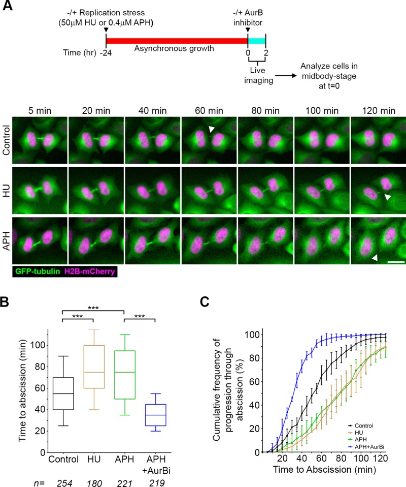
FIGURE 3:
Replication stress leads to prolonged time to abscission. (A) Timeline of experimental procedure and montages of HeLa cells stably expressing GFP-tubulin (green) and histone H2B-mCherry (magenta) cultured in HU or APH. Scale bar, 20 μm. (B) Quantification of abscission timing using the assay described in Figure 2. ***p < 0.0001. (C) Cumulative frequency plots of progression through abscission. Each point represents the percentage of all midbody cells analyzed in B that had proceeded through abscission over the time course of the experiment. Error bars are mean and SD from three to five experiments.

