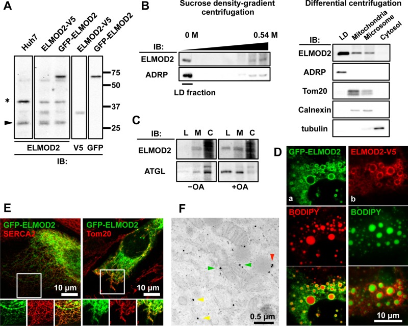
FIGURE 1:
Subcellular distribution of ELMOD2. (A) Validation of the rabbit anti-ELMOD2 antibody by Western blotting. The antibody recognized endogenous ELMOD2 in Huh7 cells. The positive reaction was observed at the mobility expected from the molecular mass of 35 kDa (arrowhead). ELMOD2-V5 and GFP-ELMOD2 expressed by cDNA transfection were also recognized by the anti-ELMOD2 antibody as extra bands corresponding to the size of the tagged protein, which was confirmed using anti-V5 and anti-GFP antibodies, respectively. A nonspecific reaction is shown by an asterisk (*). (B) Endogenous ELMOD2 in Huh7 cells was enriched in the LD fraction. Subcellular fractions of Huh7 cells were obtained either by sucrose density–gradient ultracentrifugation or by differential centrifugation. With either method, LDs were concentrated in the fraction of the lowest density, which was confirmed by the enrichment of ADRP (perilipin2). Note that ELMOD2 was also detected in the microsomal and mitochondrial fractions obtained by differential ultracentrifugation. (C) Endogenous ELMOD2 in HeLa cells was also detected in the LD fraction. Fractions enriched with LDs (L), membranes (M), and the cytosol (C) were obtained by OptiPrep density–gradient centrifugation and subjected to Western blotting with anti-ELMOD2 antibody. HeLa cells harbor few LDs in the normal culture condition (−OA), and the reaction in the LD fraction was apparent only when cells were cultured with 0.4 mM OA for 12 h (+OA). The cytosol fraction was overloaded to show a paucity of soluble ELMOD2. (D) ELMOD2, either conjugated with GFP at the N-terminus (a, green) or tagged with V5/His at the C-terminus (b, red), showed concentration around LDs in Huh7 cells. LDs were stained with BODIPY558/568-C12 (a, red) or BODIPY493/503 (b, green). (E) GFP-ELMOD2 (green) showed colocalization with SERCA2 and Tom20 (red), indicating distribution in the ER and mitochondria, respectively. HeLa cells were cultured with 0.4 mM OA for 3 h to induce LD formation. (F) Preembedding immunoelectron microscopy of GFP-ELMOD2 in HeLa cells cultured with 0.4 mM OA for 3 h. The results confirmed the presence of GFP-ELMOD2 in LDs (red arrowhead), the ER (yellow arrowheads), and mitochondria (green arrowheads).

