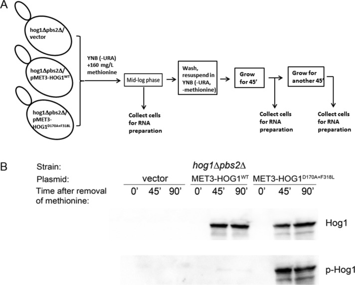
FIGURE 1:
The experimental system. Induced expression of intrinsically active Hog1 in hog1∆pbs2∆ cells. (A) Schematic description of the experimental setup. Cells of the indicated three strains were grown to mid log phase in medium containing 160 mg/l methionine, which suppresses the expression of ectopic Hog1. The cells were then washed, resuspended in medium lacking methionine, and allowed to continue proliferating. mRNA samples were collected before removal of methionine (time point 0) and at 45 and 90 min after removal of methionine. (B) Hog1 molecules were monitored 45 and 90 min after removal of methionine, and Hog1D170A+F318L was spontaneously phosphorylated. Protein lysates were prepared from cells collected at the time points at which RNA was isolated (A) and analyzed by Western blot using anti-Hog1 antibodies (top) and anti–phospho-p38 antibodies (bottom).

