FIG. 1.
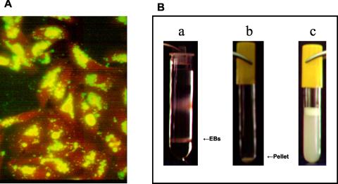
(A) Epifluorescence micrograph of HEp-2 cell monolayer infected with C. pneumoniae and stained with FITC-labeled anti-Chlamydia antibody (Pathfinder; Bio-Rad Laboratories) at 48 hpi. Greater than 95% of the monolayer contains C. pneumoniae inclusions, a level that is considered sufficient for continuation of the propagation and purification process (see detailed protocol in text). Magnification, ×340. (B) Renografin gradient containing the C. pneumoniae EB layer at the 20%-50% interphase (a); pellet of C. pneumoniae EB following a washing step (b); final suspension of C. pneumoniae EBs following trituration (c).

