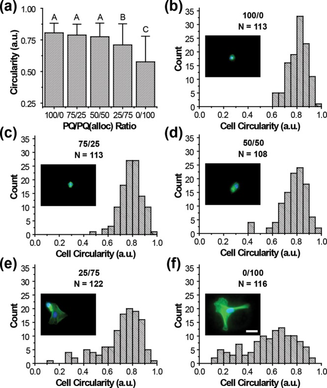
Figure 5.

Effects of hydrogel compliance on EC spreading. (a) Average circularity values for cells encapsulated in 5% PEG hydrogels with ratios of 100/0, 75/25, 50/50, 25/75, or 0/100 PEG–PQ/PEGPQ(alloc). Cell spreading, as assessed by lower circularity, increased as the hydrogels became more compliant. 25/75 and 0/100 PEG–PQ/PEG–PQ(alloc) exhibited significantly lower circularities than the other formulations and ECs in the 100% PEG–PQ(alloc) hydrogel were the most spread (p < 0.005). (b-f) Histograms that summarize the measured circularity for all the cells assessed in each hydrogel formulation. While a population of highly rounded cells (circularity ∼0.8) can be found in all hydrogels, populations of spread cells begin to emerge as the compliance decreases. Nuclear (DAPI, blue) and actin (green) staining of characteristic cells show more developed actin networks with identifiable stress fibers in highly spread cells as compared to diffuse bands surrounding the nucleus in rounded cells (inset). Scale bar represents 30 μm.
