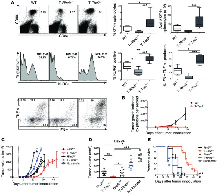
Figure 4. The ability of hyperactive mTORC1 signaling to promote effector function is cell intrinsic.
(A) OT-I+CD90.1+CD8+ T cells from WT, T-Rheb–/–, and T-Tsc2–/– mice were adoptively transferred into WT CD90.2+ recipient mice infected with vaccinia-OVA. Six days after infection, splenocytes were harvested. The percentage of splenic CD8+ CD90.1+ T cells, KLRG1 expression of CD8+CD90.1+ cells, and IFN-γ and TNF-α production after SIINFEKL peptide stimulation are shown, with statistics to the right (n = 10). For the box-and-whiskers plots, the whiskers represent the minimum and maximum values, the box boundaries represent the 25th and 75th percentiles, and the middle line is the median value. (B) WT and T-Tsc2–/– mice received s.q. EL4 thymoma cells expressing luciferase. Tumor burden was assessed by detection of luminescence (n = 16). (C–E) In vitro–activated T-Rheb–/– and T-Tsc2–/– and littermate control OT-I+CD8+ T cells were injected into WT recipients that had received B16-OVA cells 6 days prior (dashed line represents T cell transfer). “No transfer” indicates that mice did not receive OT-I+ cells. (C) Tumor volume was assessed every 2 to 3 days. Each symbol represents an average per genotype (n = 15). (D) Tumor volume shown at day 24 after tumor inoculation. Each dot represents a mouse. Data are derived from C. (E) Survival was assessed. Mice that received T-Tsc2–/– cells had enhanced survival compared with all other treatments, as determined by multiple comparisons Mantel-Cox tests. Statistics in A and D were determined by ANOVA and for B and C were determined by repeated-measures analysis. Data are representative of at least 3 independent experiments. *P < 0.05, **P < 0.01, ***P < 0.001

