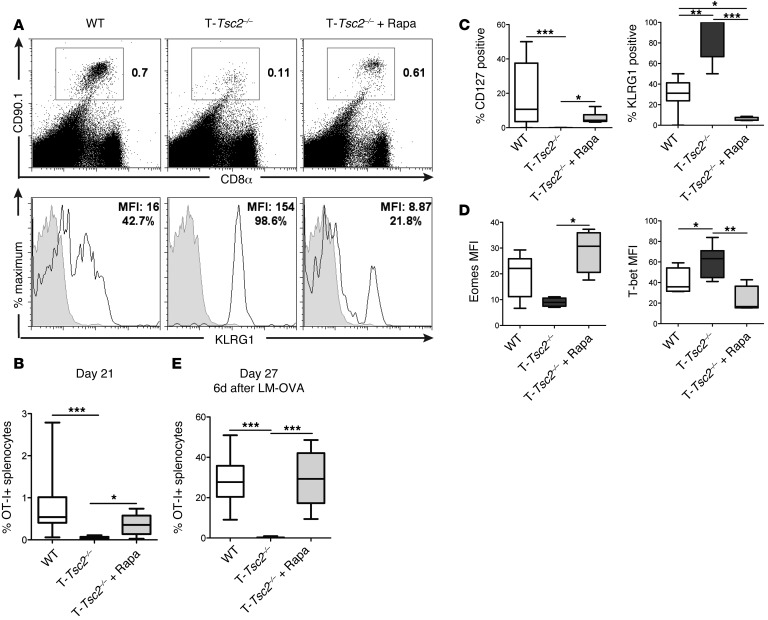
Figure 6. Rapamycin treatment can rescue the terminal effector differentiation of T-Tsc2–/– CD8+ T cells.
WT and T-Tsc2–/–OT-I+CD8+CD90.1+ T cells were adoptively transferred into WT CD90.2+ recipients infected with vaccinia-OVA. A cohort of mice that received T-Tsc2–/– cells was treated with rapamycin. (A) FACS plots depict the percentage of splenic CD8+CD90.1+ (OT-I+) cells 21 days after infection (top). KLRG1 expression of gated CD90.1+ cells (bottom). Gray histogram depicts isotype control. (B) The percentage of recovered OT-I+ splenocytes 21 days after infection (n = 8). (C and D) Phenotypic analysis of recovered OT-I+ splenocytes demonstrating (C) changes in surface marker expression and (D) transcription factor expression between genotypes with or without rapamycin treatment (n = 8). (E) Recipient mice were infected with lm-OVA on day 21 after transfer. Six days after secondary infection with lm-OVA (day 27), splenocytes were harvested and the percentage of OT-I+ splenocytes was determined (n = 14). Data are representative of 3 independent experiments. For the box-and-whiskers plots, the whiskers represent the minimum and maximum values, the box boundaries represent the 25th and 75th percentiles, and the middle line is the median value. *P < 0.05, **P < 0.01, ***P < 0.001, ANOVA. Eomes, eomesodermin.

