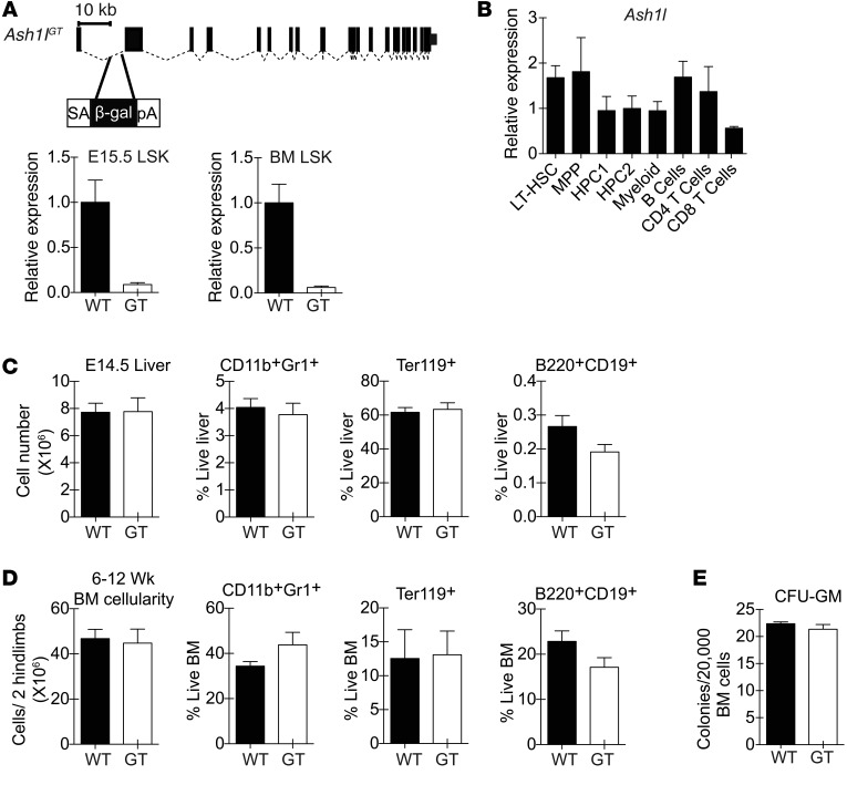
Figure 1. Preserved overall fetal and adult hematopoietic output in Ash1lGT/GT mice.
(A) Generation of the Ash1lGT allele by insertion of a splice-acceptor gene trap cassette into the first Ash1l intron. Homozygosity led to >90% reduction in wild-type transcripts in fetal (E15.5) and adult LSK progenitors, as shown by quantitative RT-PCR (qRT-PCR) with primers amplifying cDNA across the exon 1–2 boundary (mean ± SD). (B) qRT-PCR analysis of Ash1l expression normalized to Hprt1 in selected hematopoietic populations (LT-HSC, LSK CD150+CD48– LT-HSCs; MPP, LSK MPPs; HPC1, LSK CD150–CD48+ hematopoietic progenitor cells; HPC2, LSK CD150+CD48+ hematopoietic progenitor cells; myeloid, CD11b+Gr1+ myeloid cells; B cells, B220+AA4.1– B cells; CD4 T cells, TCRβ+CD4+ cells; CD8 T cells, TCRβ+CD8+ T cells) (mean ± SD). (C) Cellularity and percentage of myeloid, erythroid, and B lineage cells in E14.5 wild-type and Ash1lGT/GT (GT) fetal liver (n ≥ 4 per genotype from 2 independent experiments; mean ± SEM). (D) Cellularity and percentage of myeloid, erythroid, and B lineage cells in young adult (6- to 12-week-old) wild-type and Ash1lGT/GT BM (n ≥ 6 per genotype from >2 independent experiments; mean ± SEM). (E) Myeloid colony formation by wild-type and Ash1lGT/GT BM in CFU-GM assays (mean ± SEM, representative of 2 experiments). No statistically significant differences by t test.

