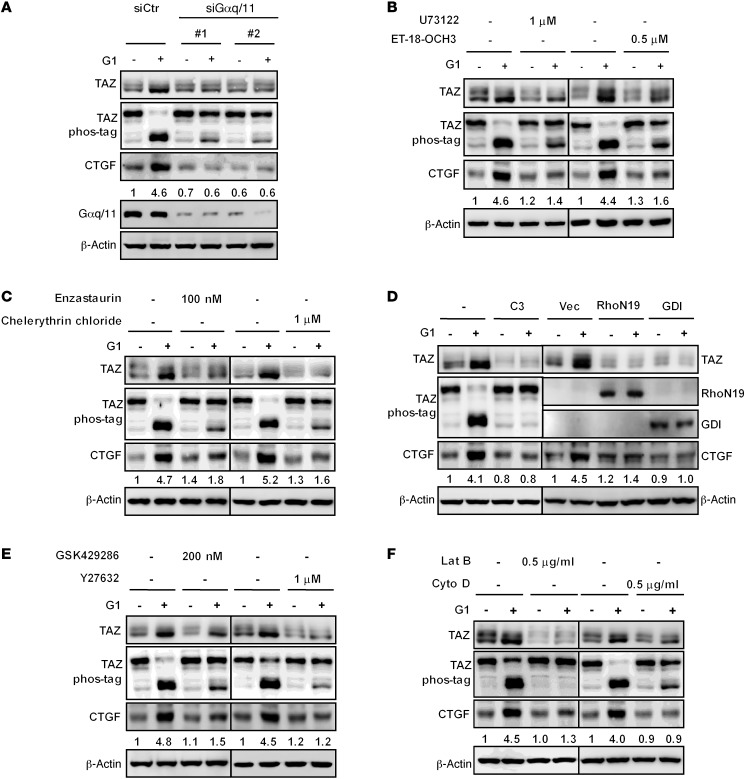
Figure 3. GPER acts through Gαq/11, PLCβ-PKC, and Rho/ROCK to stimulate TAZ.
(A) Gαq/11 was required for G1 to activate TAZ. ZR-75-30 cells were transfected with control or 2 different Gαq/11 siRNAs. After 8 hours of serum starvation, cells were treated with 100 nM G1 for 2 hours. The knockdown efficiency of Gαq/11, TAZ protein levels and phosphorylation, and CTGF expression were determined by immunoblotting. (B and C) PLCβ and PKC were required for G1-induced TAZ activation. Serum-starved ZR-75-30 cells were pretreated with PLCβ inhibitors (U73122 or ET-18-OCH3) or PKC inhibitors (enzastaurin or chelerythrin chloride) for 4 hours and then stimulated with 100 nM G1 for 2 hours. The lysates were subjected to immunoblot analysis with the indicated antibodies. (D) Inactivation of Rho prevented TAZ dephosphorylation and accumulation following G1 stimulation. Serum-starved ZR-75-30 cells were pretreated with C3 for 4 hours (left panel) or transfected with dominant-negative RhoN19 or Rho GDI (right panel) and then stimulated with 100 nM G1 for 2 hours. Cell lysates were subjected to immunoblotting with the indicated antibodies. (E) ROCK was required for G1-induced TAZ activation. After serum starvation, ZR-75-30 cells were pretreated with the ROCK inhibitor GSK429286 or Y27632 for 4 hours, followed by treatment with 100 nM G1 for 2 hours. Immunoblotting was performed. (F) Disruption of the actin cytoskeleton blocked G1-induced TAZ activation. Serum-starved ZR-75-30 cells were pretreated with Lat B or Cyto D for 15 minutes and then stimulated with 100 nM G1 for 2 hours. Immunoblotting was performed. Data are representative of at least 3 independent experiments.

