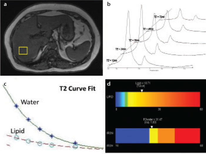
Figure 5. a–d.
The acquisition of high-speed, T2-corrected multi-echo high-speed T2-corrected MR spectroscopy involves the concatenation of five stimulated-echo acquisitions within a single 15 s breath hold. Following voxel placement (a), five echoes are acquired. Postprocessing steps automatically transform MR spectroscopy spectral signal (raw data) into visible water and lipid peaks (b). The analysis algorithm estimates the integral areas of water and total lipid, and calculates individual metabolite T2 using a nonlinear least-squares fit (c). The T2 values are used to correct the inherent water and lipid decay to produce a T2-corrected lipid fraction while also producing an estimate of iron content based on the R2 (1/T2) of the water peak. An easy to interpret color bar can be produced (d).

