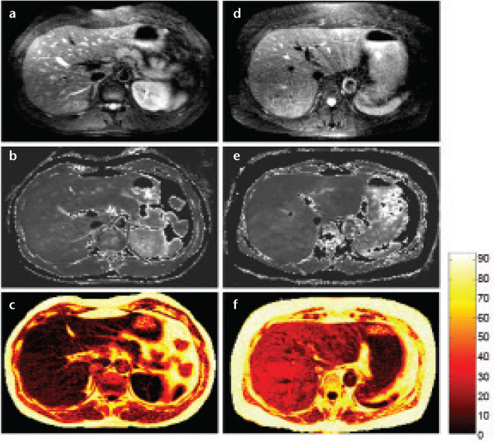
Figure 8. a–f.
Abdominal images acquired with radial-GRASE technique. Water image (a), T2-weighted map (b), and % lipid signal map (c) of a healthy volunteer. Water image (d), T2-weighted map (e), and % lipid signal map (f) of a patient diagnosed with fatty liver disease. The water image, % lipid signal, and T2-weighted maps were generated from data acquired with radial-GRASE in a single breath hold. The % lipid signal and T2-weighted maps are thresholded to null out regions that consist of mostly noise. The T2-weighted maps include lipid regions, which contain little or no water signal.

